

Clinical Staging and Classification System in Varicocele Diagnosis
Varicocele disease (a condition where the veins carrying deoxygenated blood in the scrotum enlarge and their valves dysfunction) does not progress with the same physical size or severity in every man. In order to predict the damage the disease causes to the testicle, to create a follow-up strategy, and most importantly to make the correct "surgery decision", varicocele is "graded" (Grading) by medical authorities worldwide according to its size and examination findings.
According to international standards (norms of the World Health Organization - WHO and Andrology Societies), varicocele grading is NOT determined by Scrotal Color Doppler Ultrasonography, but by physical (manual) examination performed by a specialist andrologist/urologist while the patient is standing. This distinction is crucial because the millimeter measurements written in ultrasound reports alone are not considered the gold standard in determining the patient's clinical stage, and especially whether surgery is required.
Distinction Between Clinical and Subclinical Varicocele
Before moving on to the grades, let's look at the absolute basics. Clinical Varicocele: Refers to vein enlargements that the physician can detect and touch during physical examination (either while the patient is relaxed or performing a deep breath and bearing down). Treatment decisions are made based on this. Subclinical Varicocele: Is a condition that the physician cannot feel physically, showing zero external symptoms, but which radiologists report as "having 2 mm - 2.5 mm reflux/backflow leakage" using sensitive ultrasound devices. According to international guidelines, subclinical varicocele is not operated on (except under special circumstances) and is only monitored.
Grade 1 (Stage 1) Varicocele: Mild Level
This is the mildest clinical form of the varicocele spectrum.
- Examination Finding: While the patient is standing for the testicular examination and the scrotum is in a normal relaxed position, the enlarged veins ARE NOT VISIBLE FROM THE OUTSIDE and CANNOT BE FELT BY TOUCH. However, when the physician asks the patient to take a deep breath, hold it, and bear down (Valsalva Maneuver), those internal veins (pampiniform plexus) inside the scrotum momentarily fill with blood and swell, and only then can they be felt as a cluster between the physician's fingers. As soon as the patient stops bearing down, the veins disappear again.
- Clinical Effect: It usually does not cause pain; in rare cases, a feeling of pressure may occur with prolonged standing. The probability of disrupting the semen analysis is lower compared to other grades, but it is not zero.
- Treatment Approach: If the patient is a young, unmarried man, the testicle has not shrunk, and there is no pain, he is kept under "observation" (follow-up) with semen analysis performed at 6-month intervals. However, if the patient is married, has been unable to have children for 1 year, and the varicocele has clearly (and otherwise inexplicably) impaired sperm quality, Grade 1 varicocele can also be operated on.
Grade 2 (Stage 2) Varicocele: Moderate Level
At this stage, pooling in the venous beds and valve failure have become quite pronounced.
- Examination Finding: While the patient is standing or lying down during the examination, the enlarged varicose venous cluster is NOT VISIBLE TO THE NAKED EYE. However, as soon as the physician touches the area, even if the patient does not bear down at all (without performing the Valsalva maneuver), the thickened, tortuous structure of the veins can be easily felt under the fingers (palpation).
- Clinical Effect: Since the fluid circulation and drainage balance of the testicle are disrupted, sperm damage due to increased heat (low sperm count - oligozoospermia, decreased motility - asthenozoospermia) begins to be clearly observed. The DNA fragmentation index (DFI) can often increase. It has a high potential to cause a dull ache in the groin.
- Treatment Approach: In a man with Grade 2 varicocele who has complaints of infertility and whose semen analysis results are also impaired, the "red light" has turned on for treatment. As the first option, Microsurgical Varicocelectomy surgery should definitely be performed. This is where a major increase in success for preventing infertility is achieved.
Grade 3 (Stage 3) Varicocele: Severe Level (Visible Varicocele)
This is the final and anatomically most severe stage that the disease can reach. It is the group that poses the greatest threat to testicular function and integrity.
- Examination Finding: There is no need to examine, touch, or ask the patient to bear down. The moment the patient stands and lowers their underwear, a massive, thick, dark blue/purplish, swollen, helical external vein cluster "resembling a bag of worms or spaghetti pasta" at the top of the testicle (usually on the left side), sometimes extending to the bottom of the scrotum, is CLEARLY VISIBLE TO THE NAKED EYE.
- Clinical Effect: Such intense toxic blood accumulation and scrotal temperature rise reaching up to 37 degrees Celsius cause major damage to sperm stem cells and frequently create a picture of severe infertility (severe oligospermia, with only 1-2 million sperm remaining). Unable to withstand the pressure, the testicular tissue tends to atrophy, lose volume, and shrink and soften. Very severe pain can reach a level that impacts social life and sexuality.
- Treatment Approach: Without hesitation, microsurgical intervention is required. The patient should be operated on quickly to stop the tissue degeneration and atrophy of the testicle, to prevent the decline in testosterone, and to keep the chance of having children alive. In Grade 3 patients, the post-operative surge and improvement in sperm values (if the testicle has not completely atrophied) are surprisingly high.
Why "Vein Diameter (mm) Measurements" in Ultrasound Reports Can Be Misleading
Many patients come to their appointments with ultrasound reports obtained from different centers, carrying the anxiety of: "Doctor, my ultrasound shows a reflux of 2.7 mm on the right and 3.8 mm on the left. Do I need urgent surgery?"
However, modern andrology associations do not accept "vein diameter in millimeters" measured solely by Doppler ultrasound as a surgical indication (decision for surgery). Ultrasound is only an auxiliary ("supportive") diagnostic tool in cases such as very obese patients where the abdomen obstructs the groin examination, or to confirm whether the internal anatomy carries a suspicion of a testicular tumor.
This is because, in clinical conditions, if the room is cold, the scrotum contracts and the vein is measured at 1.5 mm; if the room is warm, it relaxes and the vein diameter is measured at 3.5 mm. Depending on how much pressure the radiologist applies with the probe (transducer head) and how strongly the patient bears down at that moment (the degree of Valsalva), the vein diameter fluctuates in millimeters from test to test.
It should be noted with care that: Your treatment should be determined based on whether you can achieve pregnancy after marriage, the biology of your sperm quality, and your andrologist's GRADE 1-2-3 physical examination stage, not on your millimeter size.
Does Varicocele Grade Progress Over Time?
Yes, anatomically, varicocele has a "progressive" nature, much like a wearing vehicle engine part. After the valves are damaged, the vessel wall stretches over the years due to blood pressure, gravity, weightlifting, and other factors, losing its elasticity, and the vein clusters grow day by day.
This means that a mild varicocele starting as Grade 1 during high school years can turn into a large, visible Grade 3 varicocele by the time a man reaches his 30s and wants to marry and have children. Therefore, the statement "you have a very small varicocele, nothing will happen, go home" is medically risky. The approach of "you have a small varicocele, BUT make sure to come for a check-up with a semen analysis once a year" is the only professional vision that aligns with Andrology science and the patient's best interest.
Conclusion: The Roadmap Created by Correct Diagnosis
No matter at what stage (Grade) it is detected, varicocele is never a "hopeless problem" that should cause distress or ruin one's quality of life. Thanks to advancing Microsurgical Technology and Prof. Dr. Teoman Cem Kadioglu's 30 years of experience in specific surgical operations with a near-zero recurrence rate and high success vision, varicocele of any grade can be completely resolved with a local intervention.
If you would like to compare the grades with the pain the patient will experience or examine them in more detail, you can read our page Facts About Varicocele Pain, or if you have decided on surgery, you can read our page The Gold Standard Microsurgical Method for detailed information about how the process works.
Scientific References
This content has been prepared for medical accuracy based on international guidelines in the fields of urology and andrology and clinical studies published in peer-reviewed medical journals.
- EAU Guidelines: Salonia, A., et al. "EAU Guidelines on Sexual and Reproductive Health." European Association of Urology, 2024. [Source/Link]
- AUA/ASRM Guideline: Schlegel, P. N., et al. "Diagnosis and treatment of infertility in men: AUA/ASRM guideline." The Journal of Urology, 2021. [Source/Link]
- Scholarly Article: Liguori, G., et al. "Color Doppler ultrasound imaging in the diagnosis of varicocele." Journal of Ultrasound, 2012. [Source/Link]
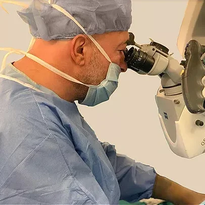
This content has been prepared by
Prof. Dr. Teoman Cem Kadioglu
Urology and Andrology Specialist